HIV Life Cycle

HIV (human immunodeficiency virus) is an infection that affects approximately 1.2 million people in the United States. The infection is very difficult to treat because the virus combines its genetic information with the genetic information of a type of white blood cell called CD4 cells. HIV can continue to replicate without treatment, generating more than 10 billion new viral particles per day.

HIV has three main stages:

  • Acute HIV. A large amount of HIV is present in your blood. Some people develop flu-like symptoms, such as chills, fever, and sore throat. It occurs about 2 to 4 weeks after exposure.
  • Chronic HIV. Once the acute symptoms subside, you enter the chronic stage where HIV replicates at low levels. You are still contagious at this stage, but you may not have symptoms.
  • AIDS. AIDS is the last stage of HIV and severely affects your immune system. It is characterized by a CD4 count of fewer than 200 cells per cubic millimetre of blood.

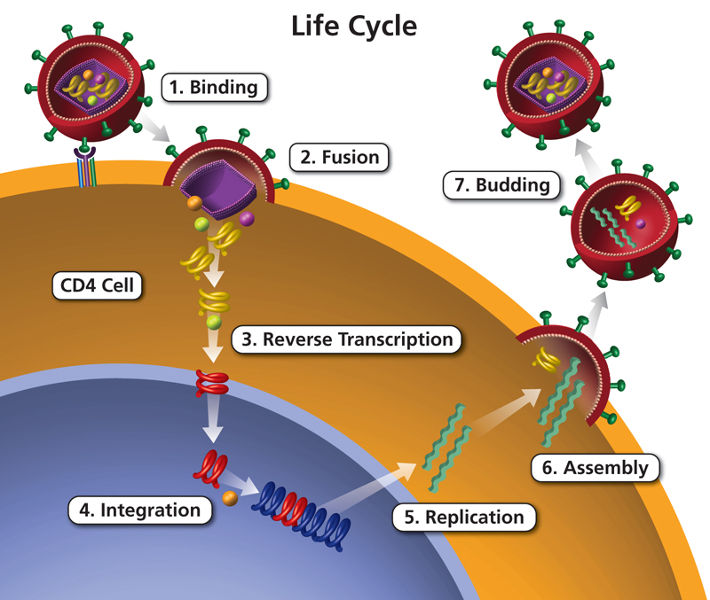
The replication cycle, or life cycle, of HIV, can be divided into seven stages. Medicines that treat HIV interrupt one of the seven stages of the HIV infection cycle. In an actively infected cell, the entire life cycle lasts only 1 or 2 days. But some cells can be latently infected, meaning that HIV can potentially be present in them for years without producing new virus particles. At any time, these cells can become activated and start producing viruses.

1. Binding

HIV belongs to a group of viruses called retroviruses. These viruses are difficult to get rid of because they integrate with the host cell’s DNA as part of their life cycle. During the first stage of the HIV life cycle, the virus binds to receptors on the surface of CD4 cells. CD4 cells, also called helper T cells, are a type of white blood cell that alert other immune cells that there is an infection in your body.

2. Fusion

HIV is an enveloped virus, which means that its genetic information is protected by both a protein layer and a lipid layer called the envelope. Once HIV binds to receptors on CD4 cells, it initiates the fusion of its envelope with the CD4 cell membrane using a glycoprotein called GP120. Glycoproteins are molecules made up of carbohydrate and protein chains. Fusion with the membrane of your CD4 cells allows the virus to enter the cell.

3. Reverse transcription

Reverse transcription is a process of converting genetic information in the form of RNA into DNA. RNA and DNA contain similar genetic information but are structurally different. RNA is usually made up of a long strand of genetic information, while DNA is made up of a double strand. The virus converts its RNA into DNA by releasing an enzyme called reverse transcriptase. This process allows the genetic information of the virus to enter the nucleus of your CD4 cell.

4. Integration

Once HIV has converted its RNA to DNA, it releases another enzyme called integrase into the nucleus of your CD4 cell. The virus uses this enzyme to combine its DNA into the DNA of your CD4 cell. At this point, the infection is still considered latent and is difficult to detect even with sensitive laboratory tests.

5. Replication

Because HIV is now embedded in the DNA of your CD4 cells, it can use that cell’s machinery to make viral proteins. During this time, it can also make more of its genetic material (RNA). These two things allow you to create more viral particles.

6. Assembly

In the assembly stage, new HIV proteins and RNA are delivered to the edge of the CD4 cell and become immature HIV. These viruses are not infectious in their current form.

7. Budding

During the budding stage, immature viruses break out of their CD4 cell. They then release an enzyme called protease that modifies the proteins of the virus and creates a mature, infectious version.

Hershey-Chase Experiment

Hershey-Chase Experiment

In 1952 (seven years after Avery’s demonstration that genes were DNA), two geneticists, A. D. Hershey-Chase Experiment provided further evidence. They worked with a DNA virus, called T2, that infects E. coli (and is also a bacteriophage). The essential element of the infectious cycle of DNA bacteriophages is T2. Virions adhere to the surface of their host cell. Capsid proteins inject the DNA core into the cell. Once inside the cell, some of the bacteriophage’s genes (the “early” genes) are transcribed (by host RNA polymerase) and translated (by host ribosomes, tRNA, etc.) to produce enzymes. they will make many copies of the phage. DNA and will deactivate (even destroy) the host’s DNA.

As new copies of the phage DNA accumulate, other genes (the “late” genes) are transcribed and translated to form the capsid proteins. The pool of DNA cores and capsid proteins assemble into complete virions. Another “late” gene is transcribed and translated into lysozyme molecules. Lysozyme attacks the peptidoglycan wall (from the inside, of course). Eventually, the cell ruptures and releases its content of virions ready to spread the infection to new host cells.

Bacteriophages produced within bacteria growing in radioactive culture medium will themselves be radioactive. If radioactive sulfur atoms (35S) are present, they will be incorporated into the protein shells of the bacteriophages, since two of the amino acids, cysteine ​​and methionine, contain sulfur. However, DNA will not be radioactive because there are no sulfur atoms in DNA. If radioactive phosphorus (32P) is used instead, DNA becomes radioactive, due to its many phosphorus atoms, but proteins do not.

Hershey and Chase found that when 32P-containing (radioactive) bacteriophages were allowed to infect nonradioactive bacteria, all infected cells became radioactive, and in fact, much of the radioactivity was passed on to the next generation of bacteriophages. However, when the bacteria were infected with 35S-labeled bacteriophages, and then the virus coats were removed (by spinning them in an electric blender), virtually no radioactivity could be detected in the infected cells. From these experiments, it became clear that the DNA component of bacteriophages is injected into the bacterial cell while the protein component remains outside. However, it is the injected component, the DNA, that can direct the formation of new virus particles complete with protein coats. So here’s one more proof that genes are DNA.

Food Spoilage

We often see that food that has been left out for too long smells bad and looks rotten. But what causes this? What will happen if we eat this food? It is eatable? Let’s find out more about food spoilage.

Introduction

If food is stored for a long period of time and is not stored properly, it will spoil; These foods are bad for your health. When food that has been stored for a long time spoils, germs begin to grow on it. Once food spoils, it cannot be eaten and must be thrown away. Spoilage is a process in which food deteriorates to the point where it is inedible for humans.

Causes of deterioration

Food and water can be infected by germs. Flies carry germs. When they sit on our food, they transmit these germs to our food. There are several factors that are responsible for food spoilage, such as bacteria, mould, yeast, moisture, light, temperature, and chemical reaction.

1. Bacteria

They are the most abundant microorganisms found on earth. They are small in size and vary in shape. Some bacteria are also helpful. They help turn milk into curds.

2. Protozoa

They are unicellular microorganisms that cause diseases such as food poisoning, etc.

3. Mushrooms

They are found in warm, moist places and grow on dead and decaying matter.

4. Temperature

Temperature is one of the main factors responsible for food spoilage.

Signs of food spoilage.

Signs of food spoilage include a different appearance than fresh food, such as a change in colour, a change in texture, an unpleasant odour or taste.

  • Activity I

Objective: To study the growth of fungi.
Materials: Piece of bread
Method: Take a piece of bread. Moisten it and store it in a warm corner of the room for 3-4 days. Observe it after 3-4 days.
Observation: Presence of greenish stain growing on the bread.

Food preservation

Food is valuable. Preserving food can help prevent food waste. Food preservation consists of preventing food from spoiling. Food preservation is the process by which food is stored by special methods. Cooked or raw foods can be preserved in different ways to be used later. Some conservation methods are:

1. Freezing

Food stored in a refrigerator stays fresh for a few days. Germs do not grow easily in cool places. We preserve foods, such as fruits with milk, vegetables, and cooked foods, by keeping them in a refrigerator.

  • Activity II

Objective: To understand the principle of food preservation.
Materials: Two apples, refrigerator.
Method: Take two apples. Keep one apple in the refrigerator and one outside for 2-3 days. Record your observation.
Remark: The apple inside the fridge is fresh while the one outside will start to rot.

2. Boiling

By this method, we can preserve food for a short period of time. Germs in milk are killed by pasteurization. It is made by sometimes boiling the milk and then rapidly cooling it.

3. Salting

We can add salt to preserve pickles and fish.

4. Sweetening

Excess sugar in food also acts as a preservative. We store food for a long time in the form of jams, jellies and murabba by adding sugar.

5. Dehydration

In this method, food is dried in the sun to stop the growth of bacteria on it. Certain foods, such as raw mangoes, fish, French fries, and potatoes, are preserved using this method.

6. Canning

In this method, the air is removed from the food and it is placed in airtight cans so that germs do not grow on it. Foods like vegetables, seafood, dairy products, etc. are preserved through this method.

Advantages and disadvantages

  • Advantages of food preservation: Germs do not grow easily on preserved foods and make them safe to eat. Conservation allows us to enjoy seasonal fruits such as strawberries and mangoes even out of season.
  • Disadvantages of food preservation: Excess salt and sugar are used in food preservation, which is not good for health. Some food preservation methods can lead to nutrient loss.